How is the procedure performed?
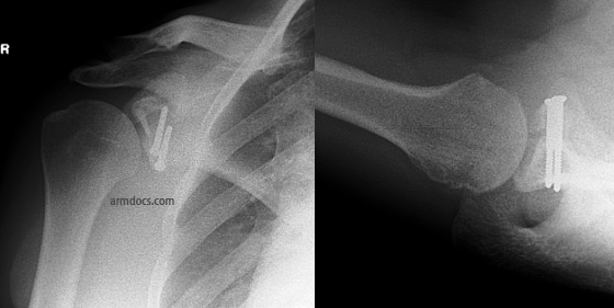
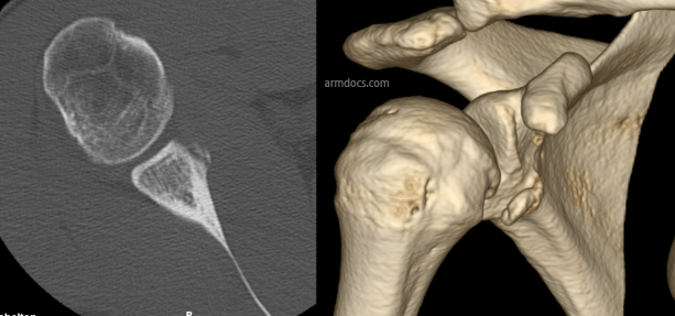
The procedure is performed under a combination of general and regional anaesthesia. The skin on the front of the joint is incised and the subscapularis tendon is split. The inside of the joint is examined. The rim of the socket is freshened to promote bleeding and facilitate healing. A bone block is prepared using the coracoid process (a part of the shoulder blade) or bone taken from the pelvis and repaired to the defect in the socket using 2 screws and occasionally a mini-plate (these procedures are referred to as the Latarjet or Eden-Hybinette procedures respectively). In selected patients the bone block may be positioned and fixed arthroscopically using a pair of suture-button devices. The labrum and capsule is fixed back to the rim of the socket with suture anchors (small devices with sutures that are embedded in bone).
Benefits
The main benefit of the procedure is to repair the damage, restore stability and improve the function of the joint. More than 90% of patients achieve benefit from surgery.
Risks
Pain in the shoulder- The shoulder may be painful for some weeks after surgery. This is usually managed by taking appropriate pain relieving medication and activity modification.
Donor site pain: If bone is taken from the pelvis, this wound is often more painful than the shoulder. You will be given pain-relieving medication. The scar on the donor site may be sensitive and some patients may develop numbness around the scar.
Bruising – Bruising is not uncommon and usually resolves over 2-3 weeks.
Stiffness – Some degree of stiffness is to be expected in the first few weeks after surgery. Prevention is the key and it is essential to follow the instructions provided by the physiotherapist and perform daily stretching within the limits imposed to maintain the range of movements. Some loss of external rotation may persist.
Graft resorption, nonunion or fracture - In some instances the graft may fail to heal or may dissolve. Fracture of the graft is rare but can occur after a further injury.
Hardware failure – Suture anchors may rarely work loose from the bone. This may require further surgery to remove the loose anchors. The screws or suture-button devices used to fix the bone block may work loose or may rarely break if the bone fails to heal.
Infection – Infection is a possibility. The risk is 1-2%. Several measures are employed to minimise the risk and you will be given antibiotics to prevent infection
Recurrence – Occasionally the bone block may fail to heal to the native socket and rarely this may result in recurrence of symptoms of instability. The risk is less than 10%.
Subscapularis insufficiency: The risk is less than 10%. This results in mild weakness of the muscle.
Nerve injury – This is possible but uncommon. The risk is about 10% and in most instances this is a minor injury that recovers over 3-4 months. Permanent nerve deficits may occur in 2% of patients.
Arthrosis – Wear and tear changes in the joint are not uncommon, develop slowly over a prolonged period of time and are usually mild.
Aftercare
Following the procedure the skin incision(s) will be closed with sutures and tape and covered with shower-proof dressings. These dressings should be left undisturbed as far as possible for 14 days. If the dressings are removed for any reason they should be replaced with similar dressings. Sutures are removed at 14 days. Prior to discharge from hospital a physiotherapist will provide instructions about looking after the shoulder. You will be advised to protect the shoulder by wearing a special sling for 3 weeks and intermittently performing limited movements of the shoulder. After 3 weeks you may stop wearing the sling during the day and will be allowed to move the shoulder actively through a greater range. You may resume driving at 4 weeks. Strengthening exercises are started after 6 weeks. Outpatient physiotherapy will be arranged and may need to be continued for 6-12 months.
Follow-up
An appointment will be arranged for you at 2-4 weeks after the procedure. Follow-up is required for at least 12 months after surgery or until a satisfactory recovery is achieved. This will include X-rays at interval to check bone healing.